

Perspectives of Targeted Radionuclide Imaging and Therapy of Fibroblast Activation Protein (FAP) in Cancer
Gourni group PD Dr. Eleni Gourni
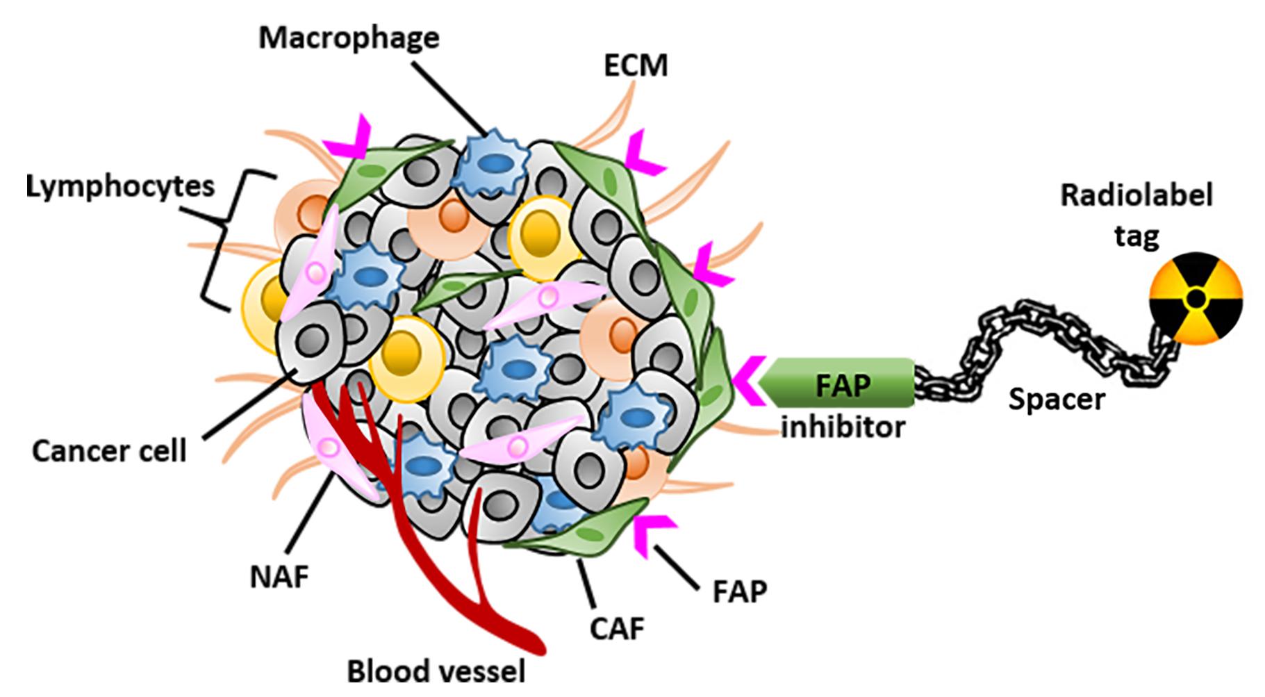
Tumors develop within a complex microenvironment consisted of diverse cell types surrounded by a matrix rich of proteins, termed tumor stroma. Stroma includes immune cells, fibroblasts and vascular enothelial cells.
Cancer cells rely on extensive support from the stroma to survive, proliferate and invade, thus making stroma an important potential target for anti-cancer therapy. Targeting elements of stroma, may be a useful therapeutic strategy to prevent tumor growth and progression. One of those elements is the fibroblast activation protein (FAP) which is overexpressed on activated fibroblasts on several tumors types.
The current project aims at designing and evaluating novel FAP-specific inhibitors for the generation of radiotracers with the potential to be used for the diagnosis and treatment of FAP-positive tumors. The novel radiotracers are thoroughly investigated in vitro and in vivo using cell lines and xenografted tumor models to understand their binding properties and their in vivo performance.
Multi-layer stratified oncology platform utilizing transcriptomics, prostate cancer organoids, and modeling of drug response
Kruithof-de Julio group Prof. Dr. phil. Marianna Kruithof-de Julio
Prostate cancer (PCa) presents a significant clinical challenge due to its multifocal and molecularly heterogeneous nature, with 60–90% of patients exhibiting multiple distinct lesions at diagnosis. Conventional treatment approaches often overlook this complexity, treating the prostate as a uniform entity and potentially missing critical lesion-specific therapeutic vulnerabilities.
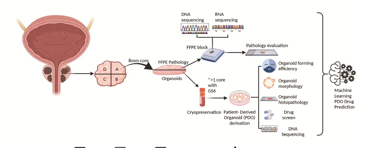
To address this, we developed a multi-layered oncology platform that integrates transcriptomic profiling, patient-derived organoids (PDOs), and functional drug screening to explore lesion-level heterogeneity and its impact on therapy response. Our objectives were to establish a twin-biopsy strategy enabling parallel molecular and functional characterization of individual lesions, generate and validate organoids from both tumor and benign tissues, identify molecular subtypes through transcriptomic clustering, and correlate these subtypes with drug sensitivity using machine learning models.
Ultimately, we aim to translate these insights into a stratified clinical workflow that supports personalized treatment decisions based on the unique molecular landscape of each patient’s disease.
Biopsies from 24 PCa patients were split into matched halves: one for histopathology and RNA sequencing, the other for organoid generation and drug screening. Organoids were successfully derived from both tumor and benign cores with a 73% success rate and characterized by immunofluorescence and genomic profiling. Transcriptomic data from FFPE samples underwent unsupervised clustering, revealing two stable molecular subtypes (C1 and C2). Drug screening involved 11 compounds, including androgen receptor and tyrosine kinase inhibitors. Machine learning models, based on pathway activity scores, were trained to predict drug sensitivity from transcriptomic data.
This integrated platform revealed that PDOs retain lineage fidelity and reflect the genomic landscape of their parental tissues. Notably, transcriptomic clustering identified subtypes with distinct drug sensitivities, C2 lesions showed heightened response to MET inhibitors like crizotinib and ponatinib, correlating with elevated MET phosphorylation.
The machine learning model reliably predicted drug response and stratified patients based on molecular subtype, with external validation from TCGA data supporting its clinical relevance. The proposed workflow combines molecular diagnostics with functional validation, enabling personalized treatment decisions even when organoid generation is not feasible.
Future directions include expanding to larger cohorts, incorporating imaging-guided biopsies, and initiating clinical trials to validate the platform’s utility in real-world settings.
Targeting Metabolic Supercomplexes in Therapy-Resistant Prostate Cancer
Pandey group Prof. Dr. phil. Amit V. Pandey
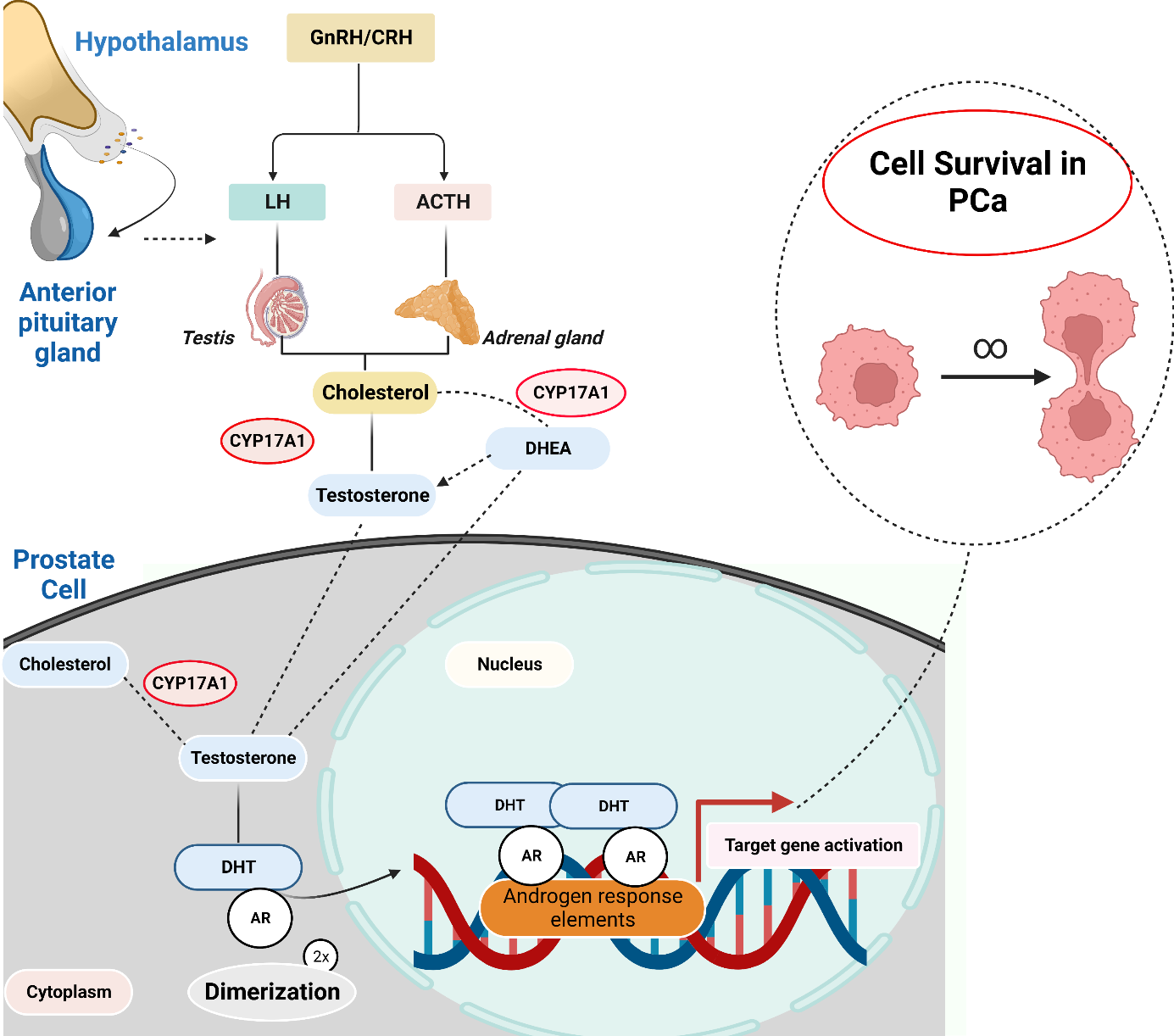
Castration-resistant prostate cancer (CRPC) represents a lethal stage of the disease, primarily driven by the tumor's ability to overcome therapy through the synthesis of its own androgens.
Our research has advanced beyond studying single enzymes to investigate their higher-order organization into what we term "metabolic supercomplexes" or "metabolons."
Our central hypothesis is that key enzymes in androgen production, such as CYP17A1, AKR1C3, and STS, do not function in isolation. Instead, they form organized, multi-protein complexes at the interface of cellular compartments, like the endoplasmic reticulum and the cytosol. These supercomplexes act as hyper-efficient production lines, utilizing a mechanism called "substrate channeling" to rapidly convert precursors into potent androgens that fuel cancer growth.
This model provides a powerful new explanation for the robust resistance observed against drugs like abiraterone. Our current work focuses on characterizing the structure and function of these supercomplexes. The ultimate goal is to develop innovative therapeutic strategies that not only inhibit key enzymes but also disrupt the crucial protein-protein interactions that hold these metabolic machines together, potentially using novel small molecules or advanced nanoparticle-based delivery systems.
Towards understanding the role of the minor spliceosome in cancer
Rubin group Prof. Mark A. Rubin MD
Genes are composed of coding units (exons), interspersed with non-coding regions called introns. The process of protein production involves splicing together exons while removing introns from the mRNA molecule. Evolution has given rise to a cellular apparatus called the spliceosome, responsible for carrying out this splicing process.
Alternative splicing enables the generation of diverse protein isoforms from a single gene. Splicing is tightly regulated under normal physiological conditions.
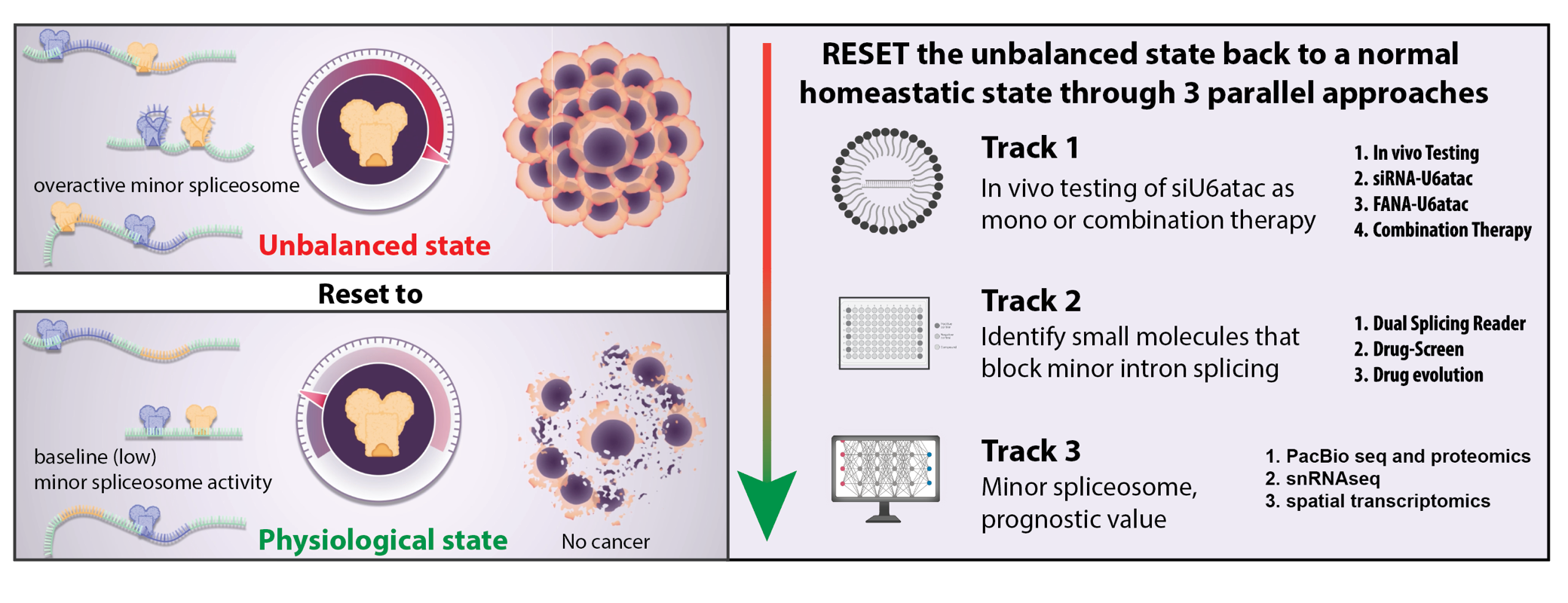
Our recent findings indicate that cancer cells use a specialized spliceosome, the so-called minor spliceosome, to increase cancer relevant mRNAs. As such cancer hijacks the minor intron-splicing machinery to enhance the expression of transcripts containing minor introns. Proteins encoded by those genes have been shown to activate critical cell survival pathways such as cell cycle regulation and DNA repair.
Exploiting the reliance of cancer cells on minor intron-containing genes presents a novel therapeutic opportunity for targeting cancer. By inhibiting the minor spliceosome, we can selectively induce cell death in cancer cells while sparing healthy neighboring cells.